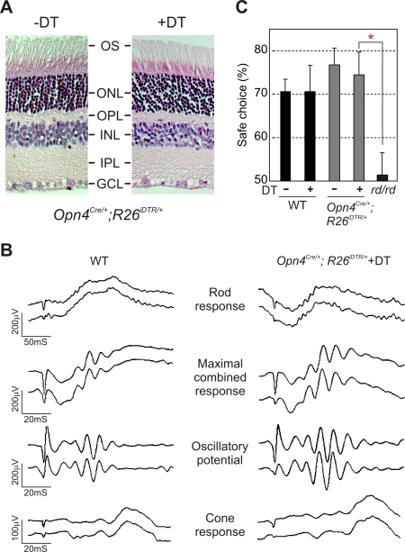
Figure 3. mRGC ablation does not alter the normal retina architecture and image-forming responses.
(A) Hematoxylin and Eosin staining of 5 μm thick paraffin embedded sections of retina from Opn4Cre/+;R26iDTR/+ mice without and with DT injection. DT application had no detectable adverse effect on the normal stratification of the retina (outer segment (OS), outer nuclear layer (ONL), outer plexiform layer (OPL), inner nuclear layer (INL), inner plexiform layer (IPL), and ganglion cell layer (GCL)). (B) Representative full-field ERG of WT and DT-treated Opn4Cre/+;R26iDTR/+ mice showing rod, cone and maximal combined responses. Responses from both eyes were simultaneously measured and plotted. Quantitative analysis of magnitude and timing of a-wave, b-wave and oscillatory potentials of these two genotype groups (3 mice each) showed no significant difference (data not shown). (C) Image forming visual function as assessed by the visual cliff test was unaffected by mRGC ablation. Average percentage (+SEM, n = 5 to 13 mice) of positive choice in 10 trials for each mouse are shown. Mice with outer retina degeneration (rd/rd) made random choices while stepping down from the platform and were significantly different (Student's t test, p<0.05; red asterisk) from the other four groups. No significant difference in test performance was found among native or DT-treated WT or Opn4Cre/+;R26iDTR/+ mice.

