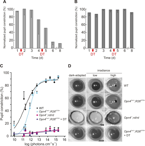
Figure 4. Necessity of mRGCs for PLR.
DT injection severely attenuates pupil constriction in response to 20 μW of monochromatic blue light (470 nm) in Opn4Cre/+;R26iDTR/+ mice (A), but does not affect in such PLR response in WT mice (B). Normalized pupil constriction (-SD; n = 3 mice) measured one day prior to or every day following DT injection for up to 8 days are shown. There was variability in the rate of loss in PLR response among Opn4Cre/+;R26iDTR/+ as reflected in the larger error bars. (C) Pupil constriction in response to varying irradiance levels over 5 log units shows the necessity of mRGC for PLR. Average (+SEM, n = 5 to 6 mice) and fitted sigmoid curves for WT, Opn4Cre/+;R26iDTR/+, Opn4−/−;rd/rd and DT-treated Opn4Cre/+;R26iDTR/+ mice are shown. (D) Representative frozen video images showing dark adapted pupil and pupil under low (1011 photons.cm−2.s−1) or high intensity light (1015 photons.cm−2.s−1) of 470 nm are shown. Notice the complete lack of pupil constriction in Opn4−/−;rd/rd and in DT-treated Opn4Cre/+;R26iDTR/+ mice. For each genotype representative images of the same eye under three different conditions are shown.

