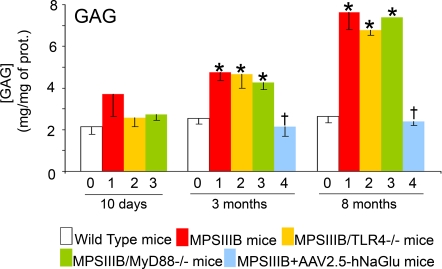
Figure 2. GAG accumulate in the brain of MPSIIIB mice.
Wild type mice (0, white bars), MPSIIIB mice (1, red bars), MPSIIIB×TLR4−/− mice (2, yellow bars), MPSIIIB×MyD88−/− mice (3, green bars), or MPSIIIB mice in which the genetic defect was corrected in the brain by a single intracerebral injection of AAV2.5-hNaGlu vector (4, blue bars) were analyzed at the age of 10 days, 3 months, or 8 months. GAG concentration was determined in cortical tissue extracts. Values are means±SEM. Asterisks indicate significant difference with wild type mouse values, crosses indicate significant difference with MPSIIIB values (p<0.05, Mann and Whitney test).

