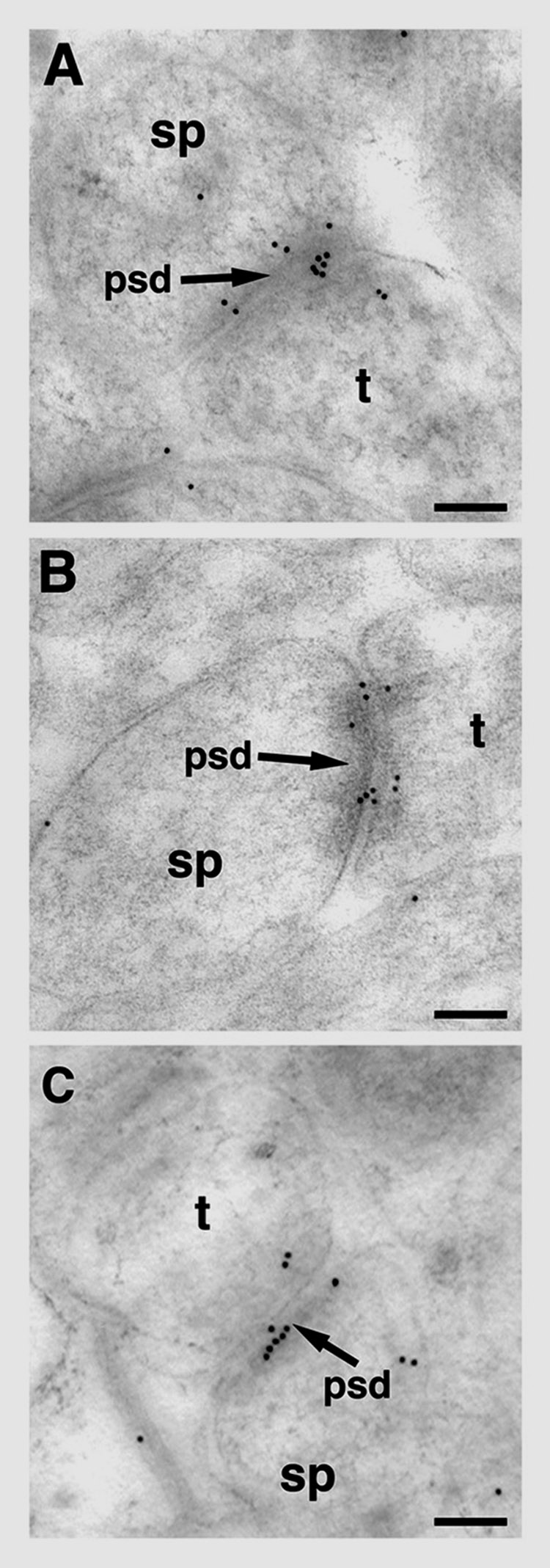
Figure 4.

Post-embedding electron microscopic distribution of pLIMK-IR within axospinous synapses in striatum radiatum of the hippocampus CA1 region. In young OVX + E female rat (A) and in young OVX + Veh female rat (B), pLIMK immunogold particles (10 nm in diameter) are seen in pre-synaptic (Bins 6, 7 and 8), cleft (Bin 5) and post-synaptic (Bins 1, 2, and 4) compartments of the synapse. pLIMK immunogold particles are affiliated with the postsynaptic density (psd) and in the post-synaptic pool (Bins 1, 2, and 4) as well as pre-synaptically (Bins 6, 7 and 8) in an aged OVX + E female rat (C). t, terminal; sp, spine; OVX, ovariectomized; E, 17-B estradiol treated; Veh, vehicle treated. Scale bars for A–C: 0.1 5m.
