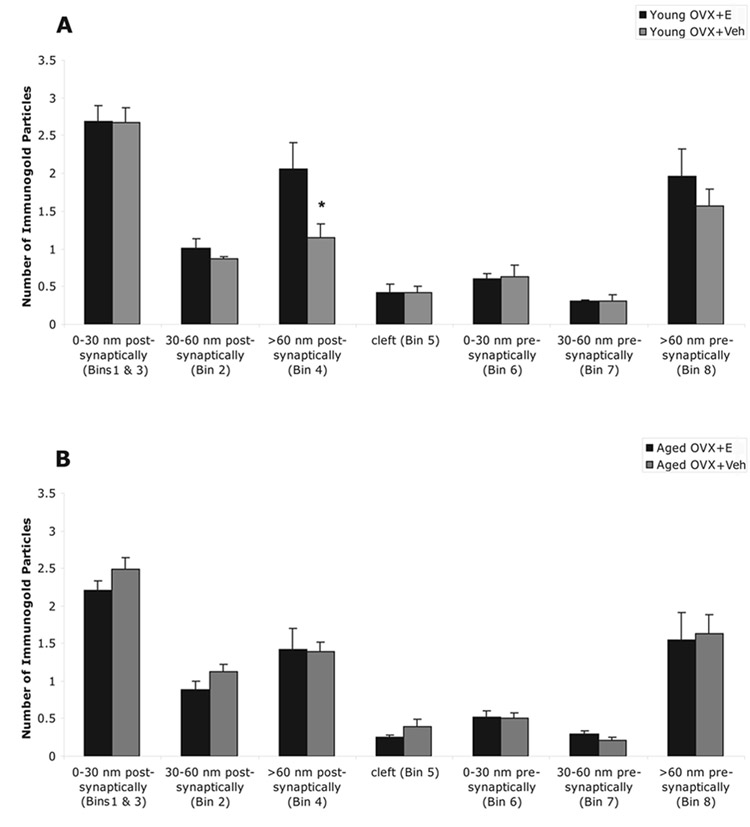
Figure 5.

Comparison of the immunogold particles in pre-synaptic, cleft and post-synaptic compartments of the synapse in four groups; A, ovariectomized (OVX) and 17β-estradiol (E) or vehicle (Veh) treated young female rats; B, OVX and E or Veh treated aged female rats. Within each group, pLIMK is mostly detected in the post-synaptic compartment. Within the post-synaptic region, the distribution of gold particles is non-uniform: highest within 0 – 30 nm (Bin 1) and 15 nm lateral (Bin 3) of the membrane associated with PSD, intermediate in the non-synaptic compartment of the spine (Bin 4) and the least in 30 – 60 nm of the membrane associated with PSD (Bin 2). (*) in A indicates E increased gold particle counts in the non-synaptic portion of the spine (> 60 nm) in young animals (p = 0.05), but not in aged animals. The age-by-treatment interaction showed a similar trend in the non-synaptic portion of the spine (> 60 nm), but failed to reach a p value of 0.05 (p = 0.08). The values are given as mean ± SEM.
