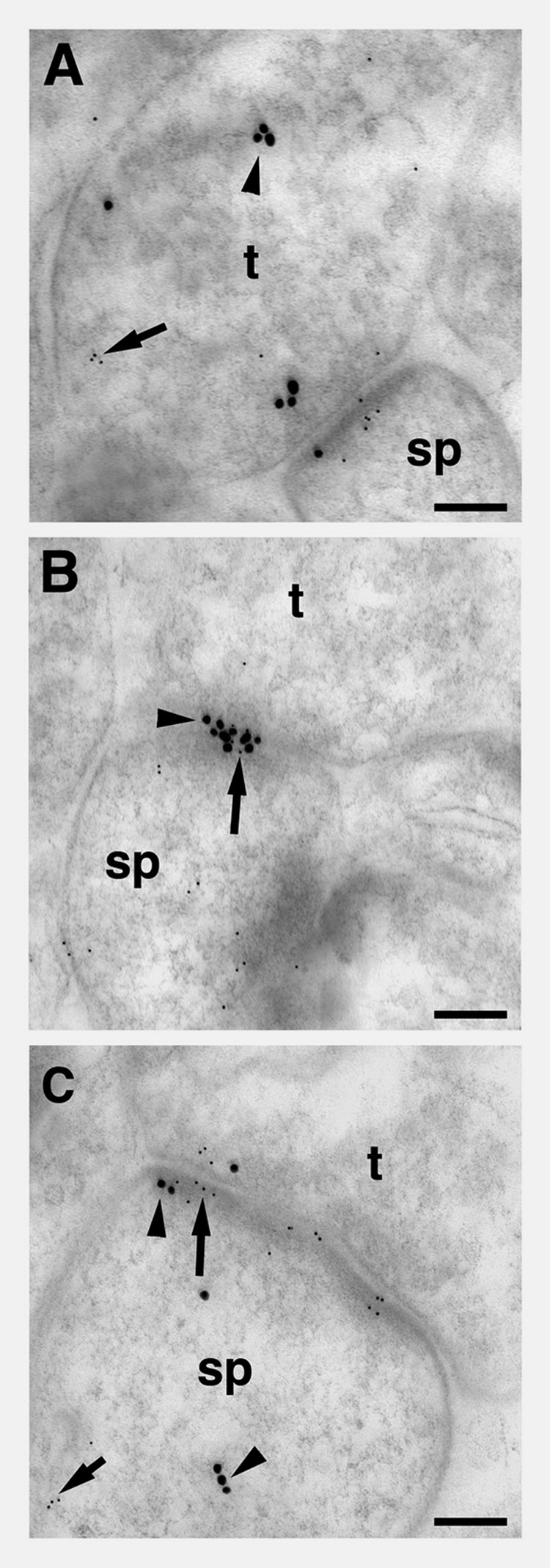
Figure 7.

Post-embedding electron microscopic distribution of pLIMK-IR relative to the ER-α-IR within axospinous profiles in striatum radiatum of the CA1 region in a young OVX + E female rat. A. In the synapse, immunoreactivities for pLIMK (5nm particles; arrow) and ER-α (15 nm particles, arrow head) in pre-synaptic compartment of the synapse (Bin 8) were observed. B. In a synapse, immunoreactivities for pLIMK (5nm particles; arrow) and ER-α (15 nm particles, arrow head) are seen in the post-synaptic density of the synapse (Bins 1, 2, 5). C. In the post-synaptic compartment of the synapse, immunoreactivities for pLIMK (5nm particles; arrow) and ER-α (15 nm particles, arrow head) are seen 15 nm lateral of the post- synaptic density (Bin 3) and in the post-synaptic compartment (Bin 4). t, terminal; sp, spine. Scale bars for A–C: 0.1 µm.
