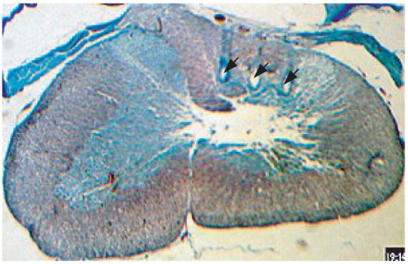
Figure 2.

Histology section from one of the experimental animals at the C5–C7 level. The section is stained using trichrome (gomori blue). The arrows point the marks left by the electrode shanks in the right half of the dorsal column.

Histology section from one of the experimental animals at the C5–C7 level. The section is stained using trichrome (gomori blue). The arrows point the marks left by the electrode shanks in the right half of the dorsal column.