
Fig. 1.
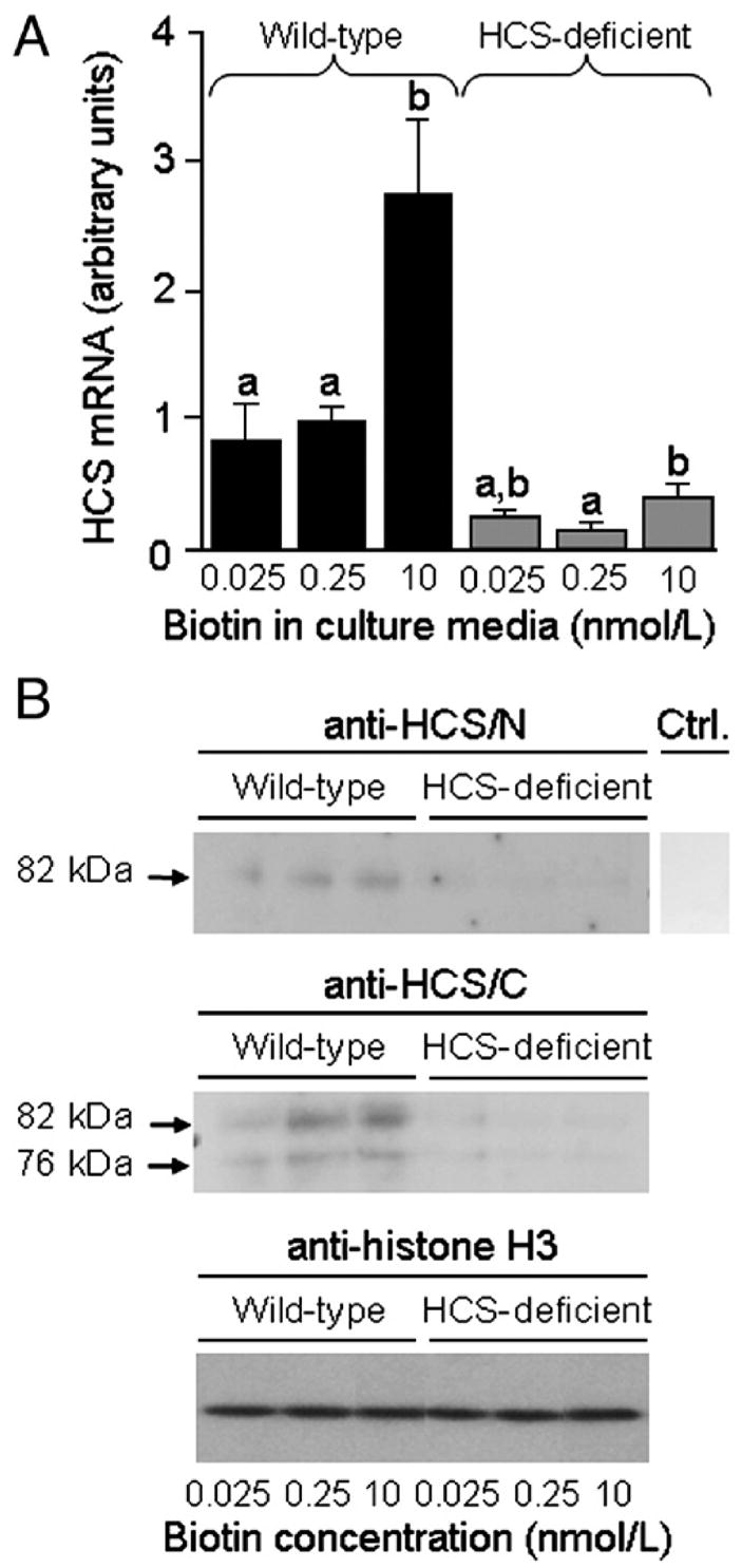
Expression of HCS decreases in response to both biotin deficiency and HCS siRNA in Jurkat cells. Wild-type cells and siRNA-treated cells were cultured in biotin-defined media for 5 weeks. (A) Abundance of HCS mRNA was quantified by real-time PCR. Values are means±S.D. a,bBars not sharing the same superscript are significantly different in cells matched for HCS expression. *P<.05, significantly different from knockdown cells matched for biotin in medium (n=3). (B) HCS protein in whole cell extracts was detected by Western blot analysis: (Top) N-terminus probed with anti-HCS/N. (Middle) C-terminus probed with anti-HCS/C. (Bottom) Equal loading of lanes was confirmed by probing blots with anti-histone H3. Ctrl. indicates control (blot probed with secondary antibody in the absence of anti-HCS/N and anti-HCS/C). Anti-HCS/N and Ctrl. samples were run on the same gel, but the Ctrl lane was processed separately after electroblotting. Samples probed with anti-histone H3 were run on the same gel, but the sequence of lanes was electronically rearranged after autoradiography.