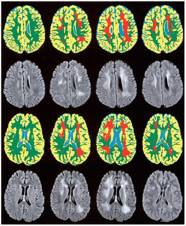
FIG. 4.
MR assessment of leukoencephalopathy in a 5-year-old girl (Subject 1, top rows) and a 10-year-old boy (Subject 2, bottom rows) treated for ALL. MR examinations (from left to right) were performed after the first, fourth, and seventh courses of high-dose methotrexate, which was administered as part of the chemotherapy regimen, and again near the end of therapy (far right). Temporal changes in leukoencephalopathy (orange) are shown on segmented and classified images of a single section. Corresponding FLAIR images are shown for reference.

