
Figure 1.
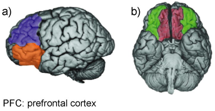
Regions of prefrontal cortex (PFC) implicated in inhibition. a) Dorsolateral PFC (blue) and ventrolateral PFC (orange). b) Ventromedial PFC (red) and orbitofrontal cortex (green). Reprinted with permission from Davidson, Pizzagalli, Nitschke, & Putman (2002). (For interpretation of the references to color in this figure legend, the reader is referred to the web version of the article.)