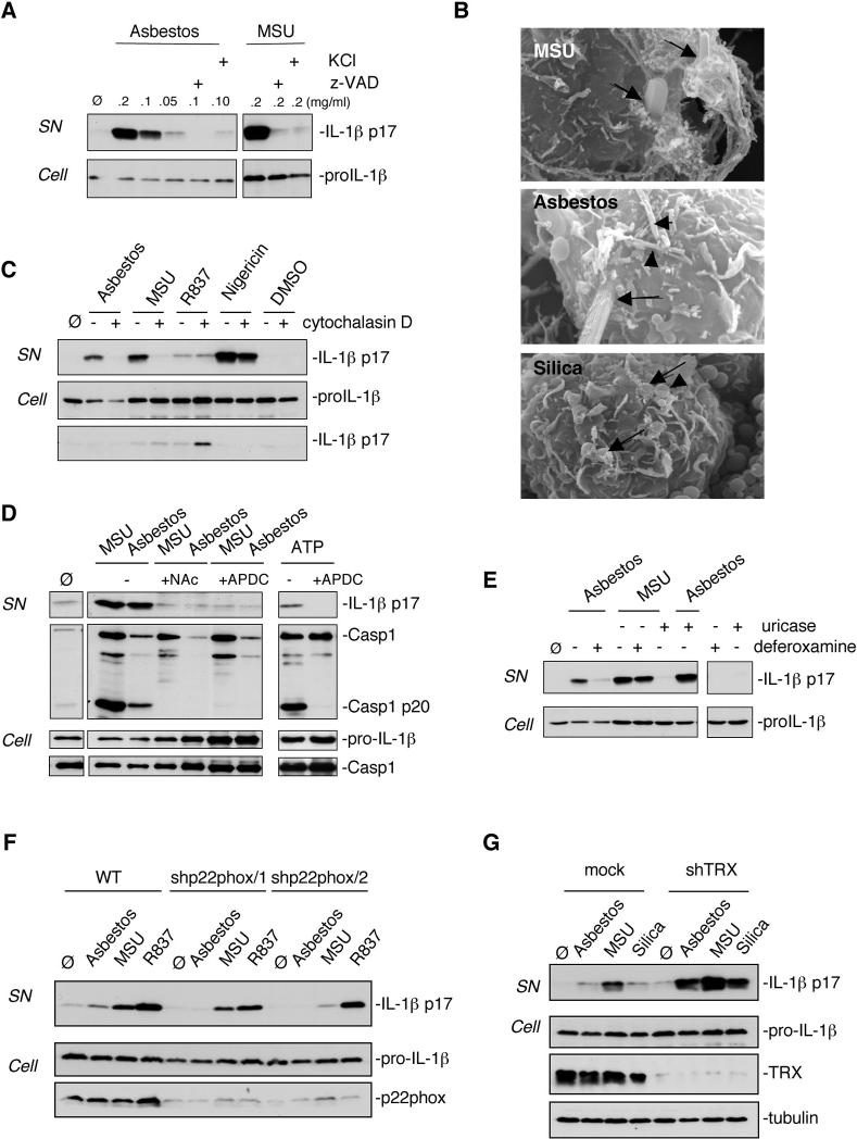
Fig 3.
Nalp3 inflammasome activation upon asbestos stimulation is dependent on endocytosis and ROS production. (A) THP1 cells were stimulated with the indicated amounts of asbestos or MSU in the presence of 20 μM caspase inhibitor zVAD or 130 mM extracellular KCl. (B) THP1 cells were treated for 6 h with 0.2 mg/ml asbestos, 0.5 mg/ml silica and 0.2 mg/ml MSU. Particles/fibers entering cells are marked with arrows and fibers/particles on cell surface with arrowheads. (C) THP1 cells were stimulated for 6 h with 0.2 mg/ml asbestos, 0.2 mg/ml MSU, 10 μg/ml R837 or 3.4 μM nigericin in the absence or presence of 0.2 μM cytochalasin D. (D) Murine peritoneal macrophages were stimulated with asbestos, MSU or ATP in the presence of N-acetyl-L-cysteine (NAC) (25 mM) or APDC (50 μM). (E) THP1 cells were stimulated with asbestos (0.1 mg/ml) or MSU (0.1 mg/ml) in the presence or absence of deferoxamine mesylate (2 mM) or uricase (0.1 U/ml) respectively. (F) THP1 control cells (mock) or knock-down for the NADPH oxidase subunit p22phox cells were stimulated for 6 h with 0.1 mg/ml asbestos, 0.1 mg/ml MSU or 0.01 mg/ml R837. (G) THP1 control cells (mock) or knock-down for thioredoxin (TRX) cells were stimulated for 6 h with 0.1 mg/ml asbestos, 0.1 mg/ml MSU or 0.5 mg/ml silica. Media supernatants (SN) or cell extracts (Cell) were analysed by Western blotting as indicated in the text.

