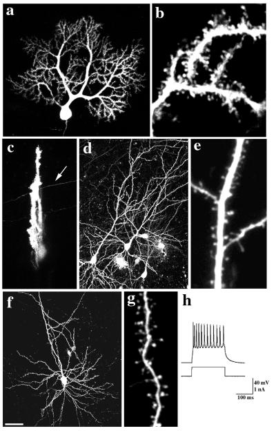
Figure 1.
GFP-transfected cells in slices have normal morphology and physiology. GFP-labeled Purkinje cells from P 10 + 2 div (a and b) sagittal or (c) frontal slices. Note labeled parallel fiber (arrow) in the frontal slice (c). (d and e) GFP-labeled hippocampal (P0 + 11 div) and (f and g) cortical (P1 + 22 div) pyramidal neurons. Individual dendritic spines on (b) Purkinje, (e) hippocampal, and (g) pyramidal neurons are clearly resolved at high magnification. (h) Whole-cell recording of action potentials elicited from a GFP-labeled cortical pyramidal neuron (10-hr acute slice) by injection of a depolarizing current. Bar = 50 μm in a, c, d, and f; 5 μm in b, e, and g.

