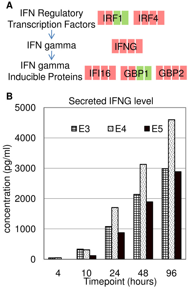
Figure 8.
Regulation of IFNG in T-cell activation. (A) Schematic showing the significantly regulated genes associated with IFNG. The regulation of gene transcription in CD3+ T cells, compared to 0 hour, is denoted by different color (green: downregulation, red: upregulation) at each timepoint in the sequence of 4, 10, 48 and 96 hours. (B) Supernatant ELISA analysis of IFNG secretion in three independent CD3+ T-cell experiments, E3–E5. CD3+ T cells were selected, stimulated (by anti-CD3/anti-CD28 antibodies) and the supernatants were harvested at the indicated timepoints of culture.

