Fig. 6.
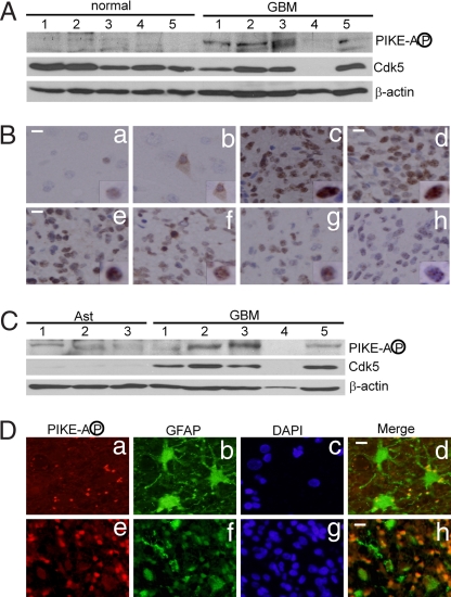
Phosphorylation of PIKE-A at S279 in human GBM tumors. (A) Levels of PIKE-A phosphorylation and Cdk5 in GBM tumors and normal human brain tissue. (B) Immunohistochemistry study of human GBM specimens. GBM tissue arrays (58 pathologically confirmed GBM cases and 6 control cases) were stained with purified anti-phospho PIKE-A antibody (Ba and Bb, normal brain tissue controls; Bc–Bh, GBM tumors). (Scale bar: 20 μm.) (C) Levels of PIKE-A phosphorylation and Cdk5 in human GBM tumors and three independent samples of normal human astrocytes (Ast). (D) Immunofluorescence studies of phospho PIKE-A in GBM tumors. GBM tissue arrays were stained with purified anti-phospho PIKE-A, anti-GFAP, and DAPI (Da–Dd, morphologically mature astrocytes in normal area of brain; De–Dh, GBM tumor cells. (Scale bar: 10 μm.)

