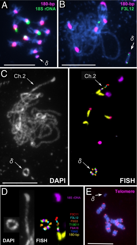
Fig. 2.
FISH images of Tr δ nuclei with mini δ (arrow) as a supernumerary chromosome. (A) A mitotic metaphase cell probed with 180-bp repeats (pink) and 18S rDNA (green). (B) Pachytene chromosomes probed with 180-bp repeats (pink) and F3L12-BAC (green). (C) DAPI and FISH images of pachytene chromosomes probed with 180-bp repeats (yellow), 18S rDNA (purple), 5S rDNA (red), and six different BAC clones; F3C11 (light red, AGI-map position on chromosome 2:1190978–1294851), F3L12 (light blue, 1288088–1404236), F5G3 (orange, 1829395–1953720), T13E11 (green, 3006966–3102924), F9A16 (deep red, 3112063–3214311), and T5M2 (deep blue, 3214009–3308310). (D) Side-by-side comparison of chromosomes 2 and mini δ; the images were derived from C. (E) A mitotic metaphase cell probed with PCR-amplified telomere DNA (5′-TTTAGGG-3′) (pink). (Scale bars: 5 μm.)

