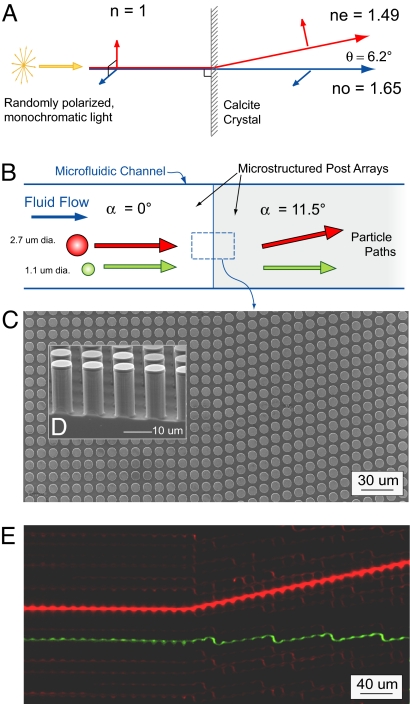
Fig. 2.
Optical and microfluidic birefringent interfaces. (A) Optical birefringence in a calcite crystal: normally incident, randomly polarized light, incident on the anisotropic crystal splits into two polarization dependent paths. Remarkably, the extraordinary ray, whose polarization is parallel to the calcite optical axis, is deflected away from the normal. (B) Schematic of particle trajectories at the interface between a neutral region and a microfluidic metamaterial element. Particles larger than a critical size follow the array asymmetry, whereas smaller particle follow the fluid flow. (C) The simplest metamaterial element is an asymmetric array of posts tilted at an angle +α relative to the channel walls and bulk fluid flow. Shown is a top-view scanning electron micrograph (SEM) of the interface between a neutral array (α = 0°) and an array with array angle α = 11.3° (the gap G = 4 μm and post pitch λ = 11 μm are the same for both sides). (D) Cross-sectional SEM image showing the microfabricated post array. (E) Equivalent microfluidic birefringence based on particle size showing the time-trace of a 2.7-μm red fluorescent transiting the interface and being deflected from the normal [see supporting information (SI) Movie S1]. Smaller, 1.1-μm red beads are not deflected at the interface; one such trace is highlighted in green to increase contrast.

