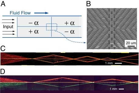
Fig. 6.
Complex microfluidic metamaterial. (A) Schematic of a complex metamaterial constructed by tiling several focusing, defocusing, and refractive elements. (B) Tilted, cross-sectional SEM image showing the interface between four subelements. (C) Collage of time-exposure images showing particle motion through a series of different +F and −F elements; motion is from left to right with just a single inlet and single outlet port. (D) A similar device with two separate inputs allowing two differently colored bead streams in the top and bottom halves of the device. Observe that particle crossover between the two halves of the device is rare; particles only mix when hydrodynamically trapped along the center reflection axis.

