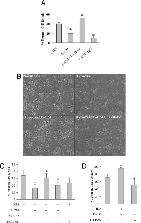
Fig. 2.
Role of BDNF in E-CM for neuroprotection. (A) E-CM protects against 24 h of hypoxia in primary rat neurons. TrkB-Fc (2 μg/ml) eliminates the neuroprotective properties of E-CM. Treatment with nonspecific IgG (2 μg/ml) has no effects. Neurotoxicity is quantified with the standard LDH release assay. (B) Representative images show primary neurons damaged by 24 h of hypoxia, rescue by E-CM, and blockade of E-CM neuroprotection with TrkB-Fc (2 μg/ml) filtering. (Magnification: 20×20) (C) E-CM neuroprotection against 24 h of hypoxia (H24) is lost when E-CM is filtered with TrkB-Fc (2 μg/ml) to remove BDNF. Addition of exogenous BDNF (10 ng/ml) restores the neuroprotective effect. Positive controls directly treated with exogenous BDNF are also protected. Neurotoxicity is quantified as LDH release from dying cells. (D) The neuroprotective capacity of conditioned media from cerebral endothelial cells is further confirmed with a cell viability assay. Hypoxia for 24 h reduced neuronal viability (MTT conversion). Treatment with E-CM provided significant neuroprotection. Filtering with TrkB-Fc to remove BDNF eliminated the neuroprotective effects of E-CM. *, P < 0.05 between hypoxic neurons alone versus neuron-endothelial cocultures or E-CM-treated neurons; #, P < 0.05 between E-CM versus TrkB-Fc-filtered E-CM conditions.

