Figure 4.
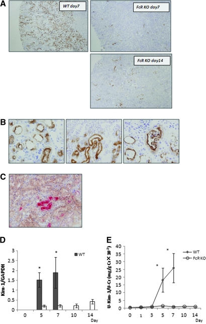
Kim-1 expression in renal tissues of WT and FcRKO mice as a sensitive marker of tubular injury. (A) Extensive Kim-1–positive tubules were seen involving almost the entire cortex of day 7 WT mice. They were predominantly found in the corticomedullary junction corresponding to S3 segment of the proximal tubules (top left). In contrast, the FcRKO mice showed minimal positive tubules at day 7 and even at day 14, despite demonstrating the comparable amount of proteinuria (top and bottom right). (B) Most tubules showed apical staining for Kim-1 staining (left). Some demonstrated dense intracytoplasmic staining of Kim-1 (middle). Occasionally, intraluminal nucleated epithelial cells were also positive for Kim-1. (right). (C) Double immunostaining showed pimonidazole-positive tubules (brown) located close to Kim-1–positive tubules (pink). Some tubules were positive for both Kim-1 and pimonidazole staining. (D) Real-time PCR revealed that Kim-1 mRNA expression in renal tissues was consistent with immunohistochemical findings (*P < 0.05, WT versus FcRKO mice on day 5 and day 7). (E) Urinary Kim-1 was elevated in WT mice correlating with protein and mRNA expression (*P < 0.05, WT versus FcRKO mice on day 5 and day 7).

