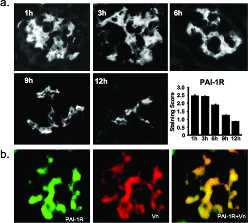
Figure 2.
(A) Time course of disappearance of injected PAI-1R from diabetic glomeruli in db/db mice at week 20. Representative photomicrographs of glomeruli from three mice at each time point that were administered an injection of PAI-1R at 100 μg/mouse intraperitoneally. A goat anti-human PAI-1 antibody (1:100; American Diagnostica) was used as the primary antibody, which was specific for human PAI-1 and did not stain mouse PAI-1. FITC-conjugated donkey anti-goat IgG (1:200 dilution; Jackson ImmunoResearch Laboratories, West Grove, PA) was applied as secondary antibody. (B) Co-localization of PAI-1R and Vn in diabetic glomeruli. A glomerulus from a db/db mouse at week 20 that was killed 3 h after PAI-1R injection. Staining for human PAI-1R (green) and mouse Vn (red). Double staining for PAI-1R and Vn (yellow). Injected PAI-1R co-localized with endogenous mouse Vn in the mesangium. Without PAI-1R injection, no staining for human PAI-1 was seen in the kidney. Magnification, ×400.

