Figure 6.
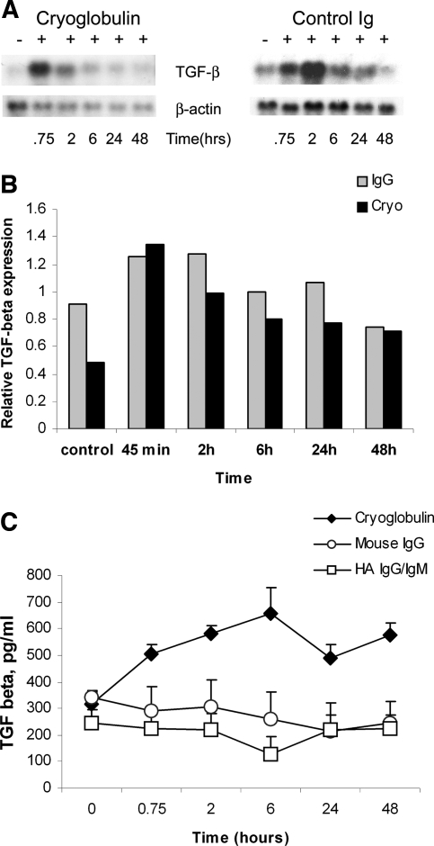
Expression of TGF-β1 mRNA and protein by mesangial cells incubated with cryoglobulin or mouse IgG. (A) Northern blot analysis. Mouse mesangial cells were incubated with cryoglobulin or control mouse IgG (control Ig) for the indicated time. Both cryoglobulin and mouse IgG induced a strong increase in TGF-β1 mRNA. Cryoglobulin induced a strong increase in TGF-β1 mRNA at 45 min, which gradually decreased up to 48 h. In contrast, mesangial cells incubated with mouse IgG showed upregulation of TGF-β1 mRNA at 45 min, which peaked at 2 h, and then decreased. (B) TGF-β1 RNA expression. There are no significant differences seen in expression levels of TGF-β1 resulting from exposure to cryoglobulins or IgG; values are normalized for equal RNA loading. (C) Analysis of conditioned medium derived from mouse mesangial cells incubated with cryoglobulin or HA IgG/IgM or mouse IgG by an ELISA specific for TGF-β1. Incubation with cryoglobulin induced increased release of TGF-β1 into culture medium as compared with control Ig. Values are the average of two experiments.

