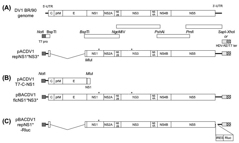
Fig.1.
(A) Schematic representation of the DV1-BR/90 genome showing the position of the restriction enzymes sites (NotI, BspTI, NgoMIV, PshAI, PmlI, SapI, XhoI and MluI) and fragments used to assemble the replicon cDNA encoded by pACDV1repNS1*NS3*. Position of the T7 promoter is shown on the left, and positions of the SapI and the HDV-RZ/bacteriophage T7 terminator fragments (which included a downstream SwaI site for linearization and an XhoI site for insertion) are shown at right. (B) Schematic representation of the structural protein-encoding cDNA fragment present in pACDV1T7-C-NS1 and the cDNA present in BAC plasmid pBACDV1flcNS1*NS3*. (C) Structure and position of Rluc encoding cistron added to the 3′ UTR of DV1repNS1*. The “*”s above NS1 and NS3 are used to indicate the position of coding differences with the BR/90 amplicon (see Table 1 and text for details)

