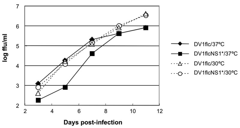
Fig.5.
Growth curves of DV1flcNS1* and DV1flc on Vero cells at 30°C or 37°C. Monolayers of Vero cells were infected with the indicated virus at a multiplicity of infection (MOI) of 0.01 and incubated at the indicated temperature. At each time point, the media were removed and frozen for subsequent titration and fresh media were added. Virus titers in cell culture medium were determined by plaque assay in Vero cells incubated at 30°C.

