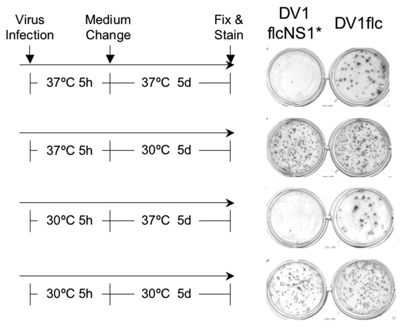
Fig.6.
Effect of temperature shift on infectious foci formation by cells infected with same numbers of ffu of DV1flcNS1* and DV1flc. The left side of the figure shows the incubation temperatures utilized for the 5-hr attachment/infection and 5-day growth/spread portion (performed under semi-solid overlay) of the experiment, and the right side of the figure shows the resulting monolayers, fixed, and stained with MAb D2-7E11 to reveal infectious foci (see Methods).

