
Figure 2.
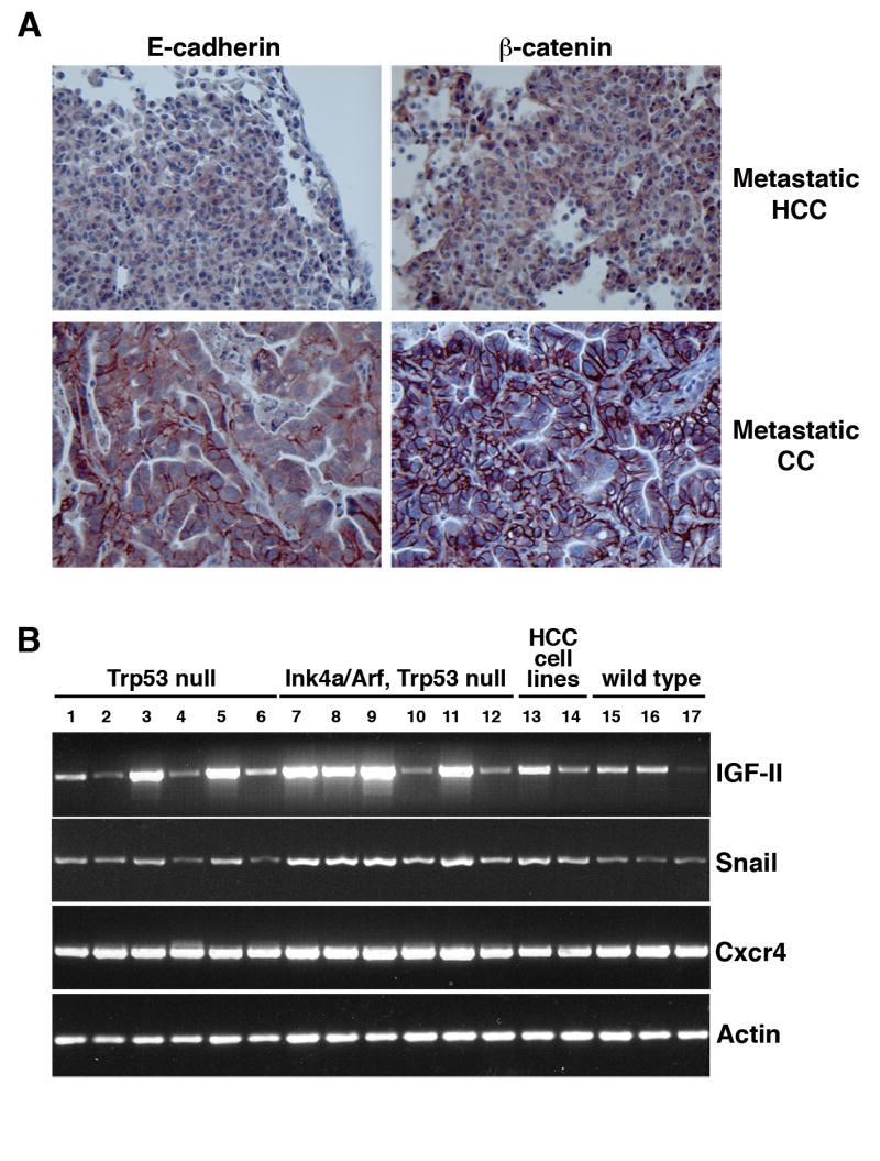
(A) Reduced expression of E-cadherin and β-catenin in an HCC-derived lung metastasis (middle panels) and retention of E-cadherin and β-catenin expression in a CC-derived lung metastasis (bottom panels) relative to a primary HCC (top panels). (B) RT-PCR analysis of wild-type, Trp53 null, and Trp53 plus Ink4a/Arf null primary liver tumors. Lanes 1-5, Trp53 null tumors; lane 6 Trp53 null GFP-infected non-tumorous liver; lanes 7-11, Trp53 plus Ink4a/Arf null tumors; lane 12, Trp53 plus Ink4a/Arf null GFP-infected non-tumorous liver; lanes 15-17, wild-type tumors; lane 13, Trp53 plus Ink4a/Arf null HCC cell line MM189; lane 14 Trp53 null cell line BL185.