Figure 7.
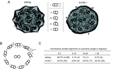
Aberrant ciliary ultrastructure in lrrc50 mutant zebrafish. (A) Electron microscopy reveals ultrastructural irregularities in lrrc50hu255H −/−. Most mutant cilia lack the ODA normally present on the outer-doublet microtubules of ciliary axonemes (I); however, misplacement of the IDA (II/III) or ODA (IV) or complete lack of all dynein arms (V) is also observed. (B) Outer-doublet alignments were measured as depicted in the schematic overview. Briefly, a best-fitting ellipse was drawn through the center (red dot 2) of the doublets. Then, a straight line (a) was drawn through the middle points of the individual doublet microtubules (dots 1 to 3). The angle between the drawn tangent line (b) through dot 2 with the best fitting ellipse and line (a) was measured. (C) lrrc50hu255H mutants show a marked increase in outer-doublet misalignments.

