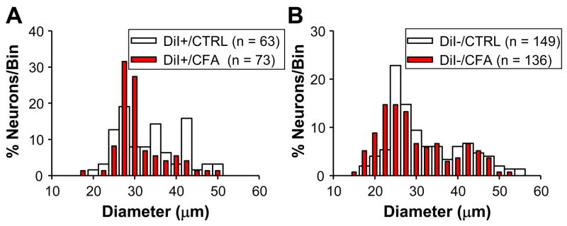
Figure 1.
Histograms of cell body diameter for naïve (CTRL) and inflamed (CFA) DiI+ (A) and DiI− (B) DRG neurons. Bin size is 5 μm. In order to facilitate comparing control and inflamed neurons, they have been plotted together and normalized with respect to the total number of neurons studied in each group. The median (+/− 25th and 75th percentiles) cell body diameter was 34.3 (29.1 and 41.0) and 32.0 (29.5 and 34.5) for DiI+ neurons from naïve and inflamed rats, respectively (p > 0.05, Mann-Whitney U test). The median cell body diameter was 30.8 (27.0 and 41.0) and 28.8.0 (25.0 and 37.5) for DiI− neurons from naïve and inflamed rats, respectively (p > 0.05, Mann-Whitney U test).

