
Figure 3.
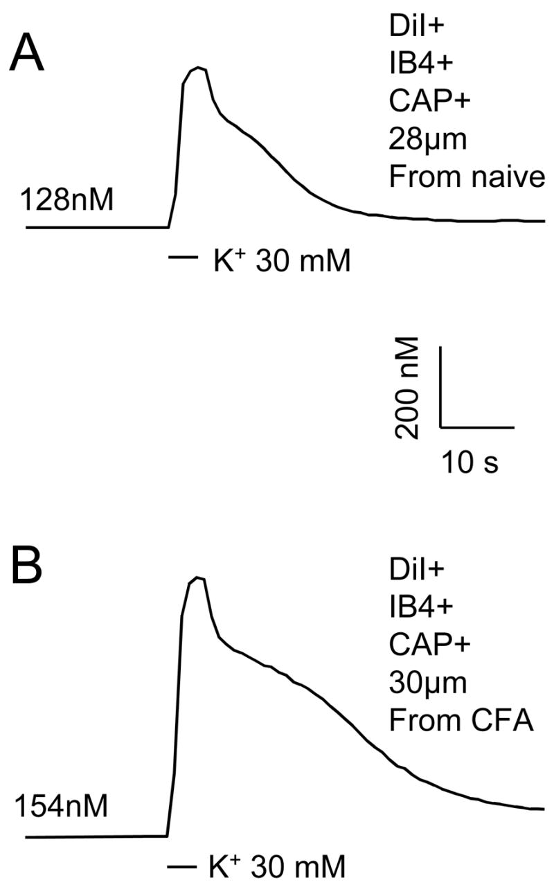
Ca2+ transients were evoked with a 4 second application of 30 mM K+ in putative nociceptive DRG neurons from a naïve (A) rat and a rat in which inflammation had been induced with a subcutaneous injection of CFA 3 days prior to harvesting ganglia (B). High K+ was applied at points indicated by black bars. Scale bars for A and B are the same. The resting [Ca2+]i in each neurons is indicated.