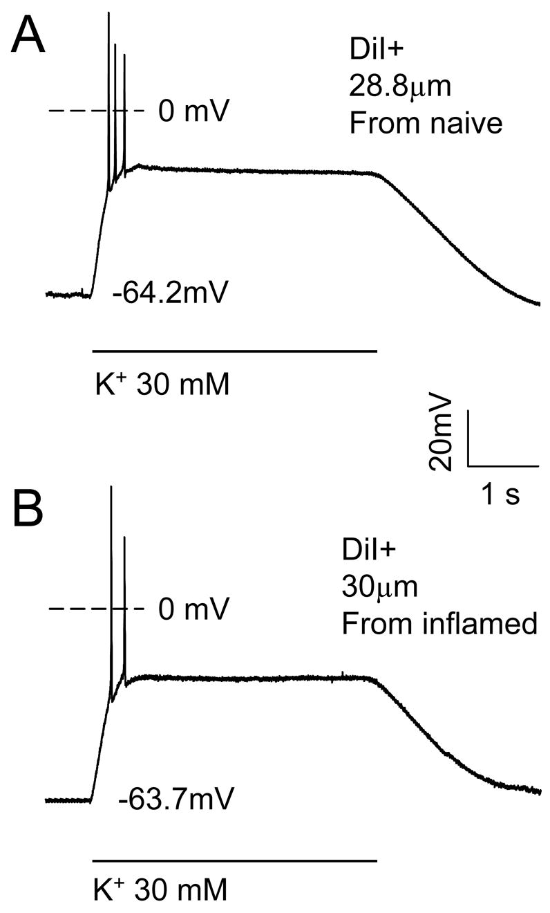
Figure 6.

Current clamp recording of the response of typical putative nociceptive DRG neurons from a naïve (top trace (28.8 μm diameter CAP responsive neuron) and inflamed (bottom trace, 30 μm diameter CAP responsive neuron) rats to 30 mM K+ (high K+). Voltage traces show a rapid depolarization that was associated with a brief burst of action potentials. The membrane potential was then stably “clamped” for the remainder of the high K+ application, after which, membrane potential returned to baseline. The resting membrane potential for each neuron is indicated.
