Figure 9.
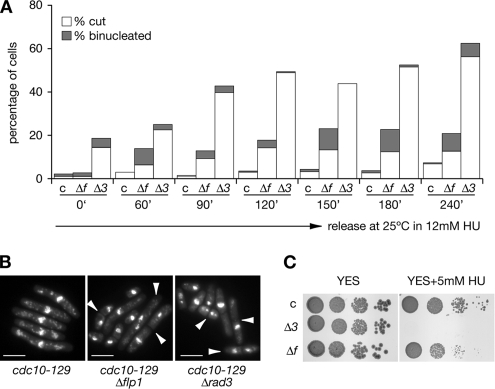
Quantification of checkpoint defects of Δflp1 mutants in synchronized cultures. (A) cdc10-129, cdc10-129 Δflp1, and cdc10-129 Δrad3 strains were synchronized in G1 (4.5 h at 36.5°C), and then they were released from the G1 block (at 25°C) in the presence of 12 mM HU. The percentages of binucleated and cut cells were estimated by counting cell samples stained with DAPI every indicated time point and plotted. At least 200 cells were counted for every strain for each time point and the experiment was repeated twice. Labels: c, cdc10-129 control; Δf, cdc10-129 Δflp1, and Δ3, cdc10-129 Δrad3 mutants. (B) Deletion of flp1+ and rad3+ causes cdc10-129 cells to accumulate binucleated and cut cells upon HU treatment. Nuclear staining (DAPI) of cdc10-129, cdc10-129 Δflp1, and cdc10-129 Δrad3 cells synchronized in G1 and released in the presence of 12 mM HU (120 min of treatment). Note the presence of binucleated and cut cells (indicated by arrows). Bar, 10 μm. (C) A 10-fold dilution plate assay showing the different sensitivity of cdc10-129, cdc10-129 Δflp1, and cdc10-129 Δrad3 strains to chronic exposure to 5 mM HU (at 32°C).

