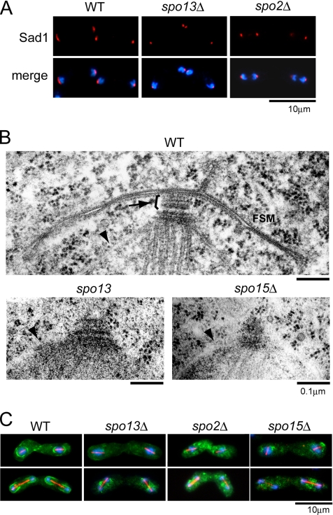
Figure 5.
Morphological changes in the SPBs and formation of the FSM in spo13Δ and spo2Δ. (A) Morphological changes in SPBs in spo13Δ and spo2Δ mutants. Wild-type (YN12), spo13Δ (YN48), and spo2Δ (YN442) strains were cultured in SSL-N sporulation medium and doubly stained with the anti-Sad1 antibody and DAPI. Blue, DAPI; red, Sad1. (B) Fine structures of SPBs in wild-type, spo13Δ, and spo15Δ strains. Wild-type (YN12), spo13 (MK13–2BL), and spo15Δ (SI52) cells cultured on SSA medium at 30°C for 1 d were observed by electron microscopy. Arrow and arrowheads indicate the meiotic outer plaque of SPB and nuclear envelopes, respectively. (C) FSM formation in spo13Δ, spo2Δ, and spo15Δ mutants. Wild-type (YN68), spo13Δ (YN66), spo2Δ (YN457), and spo15Δ (YN67) strains harboring integrated GFP-Psy1 were cultured in SSL-N sporulation medium and doubly stained with the anti-α-tubulin antibody TAT-1 and DAPI. Blue, DAPI; red, α-tubulin; green, GFP-Psy1.

