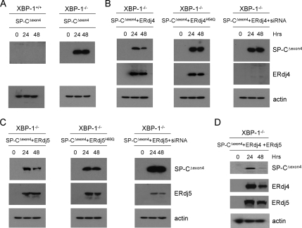
Figure 3.
ERdj4 and ERdj5 restore degradation of SP-CΔexon4 in XBP-1−/− MEFs. (A) XBP-1+/+ and XBP-1−/− MEFs were transiently transfected with plasmid encoding SP-CΔexon4 followed by Western blotting of cell lysates with SP-C and actin antibodies at 0, 24, and 48 h posttransfection. (B) XBP-1−/− MEFs were cotransfected with plasmids encoding SP-CΔexon4 and ERdj4 (left), ERdj4H54Q (middle) or ERdj4 plus ERdj4 siRNA (right). Cell lysates were prepared at 0, 24, and 48 h posttransfection, and then they were analyzed by Western blotting with SP-C, HA (ERdj4), and actin antibodies. (C) XBP-1−/− MEFs were cotransfected with plasmids encoding SP-CΔexon4 and ERdj5 (left), ERdj5H63Q (middle), or ERdj5 plus ERdj5 siRNA (right). Cell lysates were prepared at 0, 24, and 48 h posttransfection, and then they were analyzed by Western blotting with SP-C, HA (ERdj5) and actin antibodies. (D) XBP-1−/− MEFs were cotransfected with plasmids encoding SP-CΔexon4, ERdj4, and ERdj5. Cell lysates were prepared at 0, 24, and 48 h posttransfection, and then they were analyzed by Western blotting with SP-C, HA (ERdj4 and ERdj5), and actin antibodies. Within each set of experiments (A–D), samples were run on one gel to ensure identical exposure times. Actin was used to adjust exposure times across experiments.

