Figure 5.
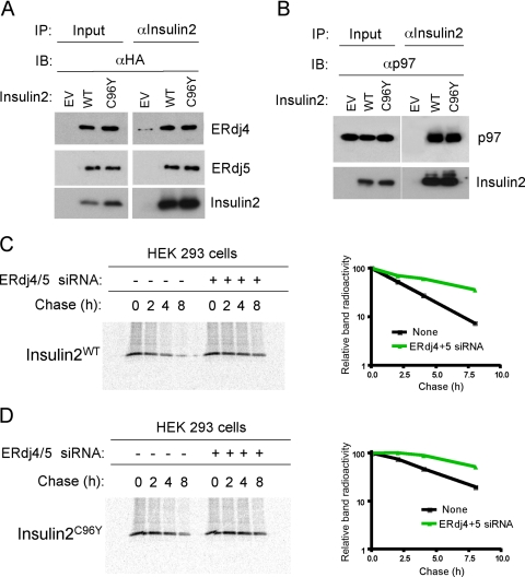
ERdj4 and ERdj5 associate with WT and mutant insulin 2. (A) HEK293 cells were transfected with empty vector (EV) or cotransfected with plasmid expressing ERdj4 or ERdj5 and plasmid encoding insulin 2 or mutant insulin 2 (insulin 2C96Y). Cells were treated with MG-132 followed by cross-linking with DSP 48 h after transfection. Cell lysates were immunoprecipitated with insulin 2 antibody followed by Western blotting with HA antibody to detect ERdj4 and ERdj5. Quantitative analyses from three independent experiments are shown in Table 2. (B) HEK293 cells were cotransfected with plasmids encoding ERdj4 or ERdj5 and insulin 2 or insulin 2C96Y. After 48 h, cells were treated with MG-132 followed by cross-linking. Cell lysates were immunoprecipitated with insulin 2 antibody followed by Western blotting with p97 antibody. (C and D) HEK293 cells were cotransfected with plasmids encoding insulin2WT (C) or insulin2C96Y (D). Pulse-chase analyses were performed as described in Figure 1B. Cell lysates were immunoprecipitated with insulin 2 antibody, subjected to SDS-PAGE, and band intensity was quantitated by phosphorimaging analyses.

