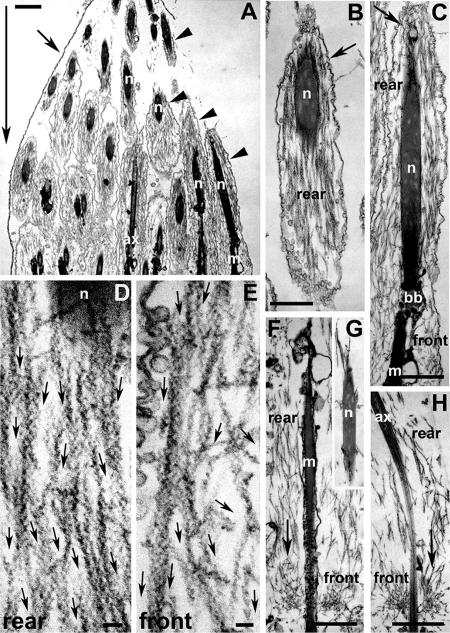
Figure 1.
Ultrastructure of S1 decorated wild-type early actin cones, before the onset of movement. (A) Longitudinal section of a single cyst terminal end (where the condensed spermatid nuclei reside). Large arrow indicates the eventual direction of cone movement. This cyst did not show any signs of cystic bulge formation. The cyst cell membrane (small arrow) can be seen enclosing some actin cones (arrowheads). (B) The rear domain of an early cone was composed of actin bundles near and around the spermatid nucleus. Most of the actin bundles were parallel to the longitudinal axis of the cone. The syncytial membrane (arrow) is indicated. (C) Longitudinal section of a whole early cone. The cone was composed of actin bundles in the rear domain (around nucleus) and short actin filaments in the front domain. Small arrow indicates the syncytial membrane. (D and E) High-magnification images of the early cone rear (D) and front (E) domains. Small arrows indicate the direction of the pointed end of each actin filament, as judged by the “arrowhead” shape generated by the myosin II S1 fragment that was used to decorate the filament. (D) The rear domain was composed of parallel-bundled F-actin with pointed ends facing in the direction of (eventual) movement. (E) Actin filaments in the front domain were less bundled, and some were oriented more perpendicular to the cyst long axis, but the pointed ends still pointed forward. (F–H) In one cyst with cones near the nuclei and no cystic bulge, a small amount of actin meshwork was visible at the front of the cones (small arrows). In the rear domain, actin bundles parallel to the longitudinal axis of the cones are visible. (G) Around a spermatid nucleus located behind an actin cone with a little meshwork, only a few actin filaments were visible; ax, axoneme; bb, basal body; m, mitochondria; n, nucleus. Bar, (A–C and F–H) 1 μm; (D and E) 0.1 μm.

