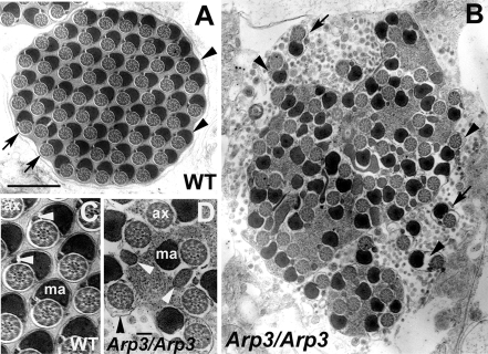
Figure 8.
Electron microscopic analysis of cross sections after individualization. (A) Each wild-type cyst contained 64 pairs of spermatid axonemes (arrows) and mitochondria derivatives (arrowheads) enclosed by a plasma membrane. (B) Individualization defects in the arp3 mutant. The arp3 mutant cyst contained only a few individualized regions (arrows). Most of the axoneme/mitochondrial pairs were not separated by plasma membrane and cytoplasm/membranous organelles were still present. The space between the axoneme/mitochondrial derivative pairs, and the membrane is larger that in wild type and sometimes contained cytoplasm and organelles (arrowheads). (C and D) Sperm tails of wild-type (C) and arp3 mutant (D) at higher magnification. Both axoneme and major mitochondria derivative structure appeared normal, but some cytoplasm remained surrounding the axoneme in arp3 mutant (black arrowhead in D). However, the morphology of the minor mitochondria derivative was abnormal in the arp3 mutant (white arrowheads in D; compare with wild-type); ax, axoneme; ma, major mitochondria derivative. Bar, (A and B) 1 μm; (C and D) 0.1 μm.

