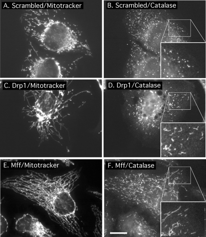
Figure 4.
Effects of Mff siRNA on peroxisomal morphology. (A) HeLa cells transfected with scrambled oligonucleotides showing wild-type mitochondrial morphologies by staining with MitoTracker. (B) The same cells stained with catalase antibody, showing the punctate distribution of peroxisomes. The inset shows an enlargement of the peroxisomal staining. (C and D) HeLa cells transfected with Drp1 siRNA oligonucleotides, showing highly connected mitochondria and elongated peroxisomes. The enlargement shows the peroxisomal defect more clearly. (E and F) Similar patterns were obtained with Mff siRNA oligonucleotides. The boxed areas, enlarged in the bottom right-hand corners of D–F, show close-ups of peroxisomes. Bar, 10 μm.

