Figure 5.
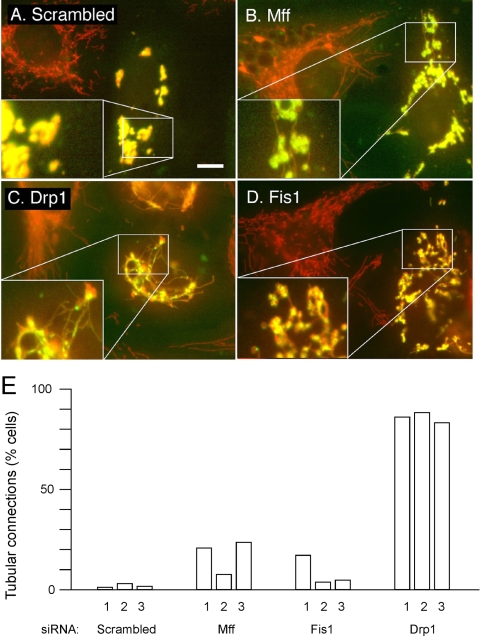
Reversal of mitochondrial fragmentation caused by dominant-negative Mitofusin expression constructs. (A) HeLa cells were transfected with scrambled Mff siRNA oligonucleotides as a control and with a myc-Mfn2(K109T) expression construct. Mitochondria were detected with MitoTracker (red). Cells expressing mutant Mfn2 were detected with myc antibody (green). These cells invariably had fragmented and often clumped mitochondria, most likely due to overexpression of aberrant mitochondrial outer membrane protein. Similar transfections were done with Mff (B), Drp1 (C), and Fis1 (D) siRNA oligonucleotides along with the myc-Mfn2(K109T) expression construct. The cotransfected cells show clumped mitochondria, but these mitochondria often have thin tubular connections, which were never detected when the myc-Mfn2(K109T) construct was transfected alone. The enlargements in the bottom left corners show these connections more clearly. Bar, 10 μm. The percentages of cells with mitochondrial connections are shown in E. The experiments numbered 1 were done with GFP tagged Mfn1(K88T), and the experiments numbered 2 and 3 were done with myc-Mfn2(K109T). For each point, 250–400 cells were counted.

