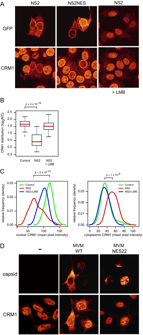
Figure 2.
NS2 relocalizes CRM1 from the nucleus to the cytoplasm (A) MCF7 cells expressing NS2-GFP (left and right) or NS2-NES-GFP (middle). When indicated, cells were treated with LMB for 3 h. Cells were stained with anti-CRM1 antibodies. (B) Boxplot summarizing quantifications (n = 36 each) of CRM1 subcellular distribution exemplified in A. Medians are indicated in red. (C) Distribution of nuclear (left) and cytoplasmic (right) CRM1 intensities, showing both an NS2-dependent nuclear decrease and a cytoplasmic increase, which is LMB-sensitive. Densities were calculated using a bandwidth of 8. p values according to Mann-Whitney tests (D) A9 cells mock-infected (−) or infected with MVMp (wt) or MVMp-NES22 (NES22) for 48 h and stained for both capsid and CRM1.

