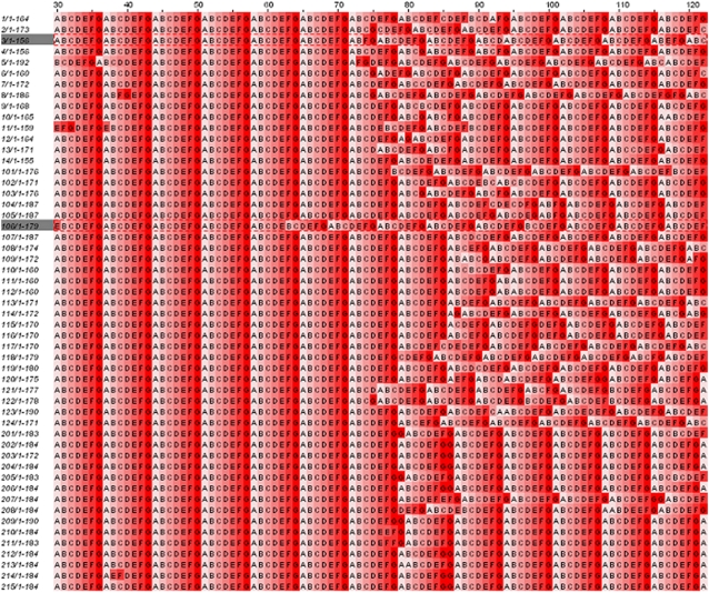
FIGURE 3.
Heptad repeats are in all compared sequences. In a different visualization using Jalview version 2.2.1 (81), the Paircoil2-assigned heptad positions of the sequences compared in Fig. 1 are shown as different shades of red. The heptad positions for E. coli amino acids are shown in the third row. The amino acid numbers for E. coli are given on top of the figure. The alignment is shown between E. coli amino acids 30 and 122, which show the highest propensity for left-handed coiled-coil formation.

