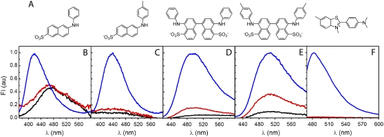
FIGURE 2.
Fluorescence probes for aggregated states of AS. (A) Chemical structures of the dyes used in this work: (left to right) 2,6-ANS, 2,6-TNS, bis-ANS, bis-TNS, and ThioT. (B–F) Fluorescence emission spectra of (B) 2,6-ANS, (C) 2,6-TNS, (D) bis-ANS (E) bis-TNS and (F) ThioT, in buffer alone (black), or in the presence of monomeric (red) or purified fibrillar (blue) AS. The curves corresponding to the dye in buffer or with monomeric AS are multiplied fivefold. Dye, 1 μM; protein, 10 μM.

