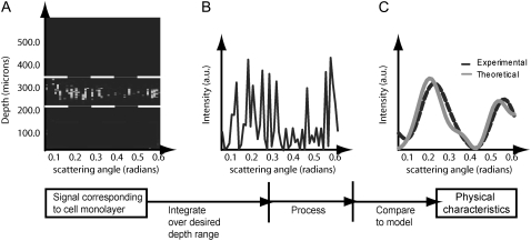
FIGURE 1.
Acquisition and processing of light scattering data from a cell monolayer using the a/LCI technique. In A, the optical depth corresponding to the cell monolayer is summed to obtain the intensity of the scattered light versus scattering angle (B). The scattering distribution is then low-pass filtered and a second-order polynomial is subtracted to isolate the nuclear scattering. Finally, the processed scattering distribution is compared to a database of known scattering distributions (calculated from an appropriate light scattering model) to ascertain the nuclear morphology (C). In this case, the sample comprised a monolayer of chondrocyte cells for which the a/LCI size determination was 6.5 μm.

