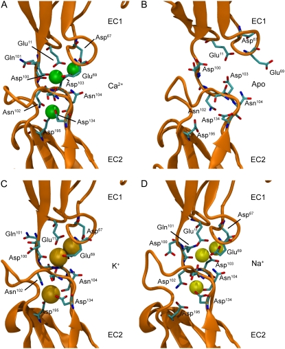
FIGURE 2.
Linker region of C-cadherin repeats EC1-EC2. (A–D) Snapshots of the linker region between repeats EC1-EC2 of C-cadherin after 1.1 ns of simulation for systems with crystallographically resolved Ca2+, without Ca2+, with K+, and with Na+ ions, respectively. The protein is shown in orange cartoon representation and ions are shown as green (Ca2+), dark yellow (K+), and light yellow (Na+) spheres. Residues originally involved in Ca2+ binding are labeled and shown for all snapshots in licorice representation.

