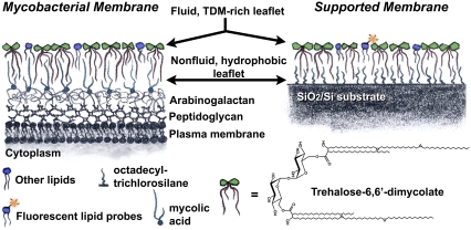
FIGURE 1.
Schematic illustration of the mycobacterial envelope and our experimental supported membrane system. In both, the outermost leaflet is a two-dimensionally fluid lipid monolayer rich in the glycolipid TDM. Underlying this outer leaflet is a dense, hydrophobic, nonfluid monolayer composed in mycobacteria of mycolic acids covalently bound to the arabinogalactan layer underneath, and in our model platform of octadecyltrichlorosilane covalently bound to a silicon wafer.

