
Figure 1.
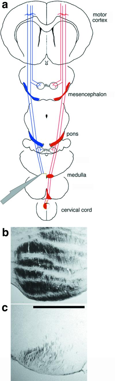
Unilateral transection of the CST. (a) CST axons arise from layer 5 pyramidal cells in the sensorimotor cortex, extend projections to the mesencephalic nucleus ruber (Ru) and pontine nuclei (PN), then assume a ventromedial position in the lower brainstem. In the caudal medulla, CST axons decussate and course in the dorsal funiculus of the contralateral spinal cord. Axons were transected via a ventral approach in the left medulla before the decussation. (b and c) Controls to verify the extent of surgery. Numerous fibers are seen in the left rostral medulla after left-sided BDA labeling (b); caudal to the transection, fewer than 10% of the original fibers remain. (c: bar = 500 μm.)