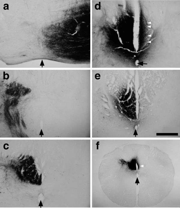
Figure 2.
Inosine induces collateral sprouting in the adult rat CST. (a–d) Axon trajectories in a case in which we transected the CST in the left medulla (before the decussation), treated the right (nonaxotomized) sensorimotor cortex with inosine for 2 weeks, then traced fiber trajectories by injecting BDA into the right sensorimotor cortex. At more rostral levels, BDA-labeled axons remain strictly lateralized and are seen in the right ventral brainstem (a), left caudal medulla (b), and left dorsal funiculus (c). At the level of the cervical enlargement, however, numerous labeled axons also appear in the denervated (right-sided) dorsal funiculus (d; white arrowheads point to individual crossed fibers). (e) Controls treated with PBS instead of inosine after unilateral CST surgery show few crossed fibers. (Bar = 200 μm.) (f) A low-magnification image of the cervical cord from a case with left CST transection, inosine treatment in the right sensorimotor cortex, and BDA labeling in the right cortex. The white arrow points to dense axon staining on the denervated side. The midline is indicated in all sections with black arrows (pointing to the central canal in c–f).

