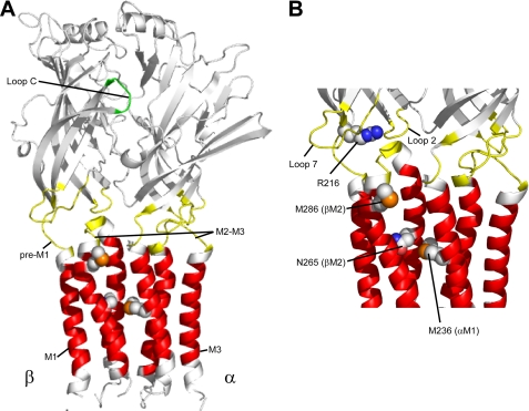
FIGURE 1.
Structural model of the GABAAR α1 and β2 subunits. A, the extracellular binding domain is colored in white. Domains believed to contribute to the GABA transduction mechanism (Loop 2, Loop 7, M2-M3 linker, and pre-M1) are highlighted in yellow. The Loop C region of the GABA binding site is highlighted in green. The transmembrane domains (M1, M2, and M3) are colored in red. Residues in the pre-M1 region (Arg-216) as well as residues forming the potential PB/general anesthetic binding site (Asn-265 and Met-286) in the β2 subunit and (Met-236) in the α1 subunit are shown in a space-filled format. The M4 transmembrane helix has been omitted for illustration purposes. B, detailed view of the interface between the ligand binding domain and the transmembrane domain.

