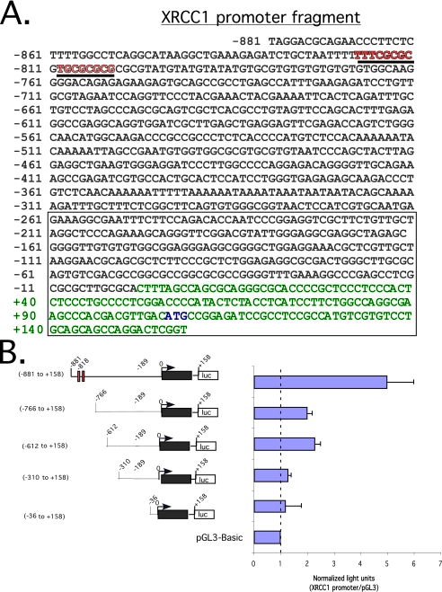
FIGURE 2.
XRCC1 promoter fragment sequence and luciferase reporter assay define an active promoter region. A, genomic sequence from -881 to +158 relative to the predicted transcription start site/cDNA 5′-end indicated as +1. Exon 1 is indicated in green type, and the translation-initiation codon is shown in blue type. The open box indicates a CpG island. Underlined sequences in red denote putative E2F-binding sites. B (left), schematic representation of a series of promoter deletion mutants in pGL3-Basic-luciferase reporter. The red boxes denote putative E2F-binding sites. The black box represents exon 1. An open box with [luc] denotes luciferase reporter. Right, luciferase readout of the indicated promoter-reporters after transfection into Saos2 cells. Light units normalized to a β-galactosidase signal. In all, 1.0 μg of the indicated promoter-reporter and pRSV β-galactosidase plasmids was used for all transfections. S.D. of triplicate experiments is shown.

