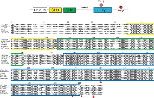
FIGURE 1.
Cloning of MbSrc1. MbSrc1 was isolated by PCR from a M. brevicollis cDNA library, as described under “Materials and Methods.” The domain structure of Src kinases is shown at the top with the regulatory tyrosines Tyr-419 and Tyr-530 (human c-Src numbering) indicated. The alignment of Src sequences shows human (c-Src Hsap), fly (Src648 Dmel), hydra (STK Hvul), sponge (Src6 Eflu), M. brevicollis (Src1 Mbre), and M. ovata (SrcFv Mova). The SH3 domain sequence is shaded yellow, the SH2 domain is green, and the catalytic domain is blue. Red circles on the sequence indicate the positions of tyrosines corresponding to Tyr-419 and Tyr-530. Arrowheads indicate residues in c-Src that are important for recognition by Csk (21); red arrowheads show residues that are conserved between c-Src and MbSrc1, and the white arrowhead shows a non-conserved residue.

