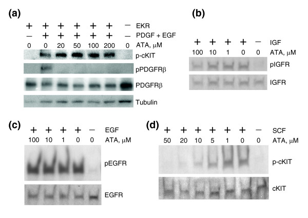
Figure 5.

ATA failed to inhibit activated EKR, IGF1R, or EGFR, but inhibited SCF-mediated activation of cKIT. (a) ATA does not inhibit activated EKR. Western analysis of total TIP5 cell lysates. Cells were transfected with EKR plasmid, serum-starved overnight and treated with ATA, EGF and PDGF. p-cKIT, pPDGFRβ, PDGFRβ, and tubulin indicate antibodies against phospho-cKIT, phospho-PDGFRβ, total PDGFRβ, and total tubulin, respectively. (b) ATA does not inhibit activated IGF1R. Western analysis of total TIP5 cell lysates. Cells were serum starved overnight and treated with ATA and IGF. pIGFR and IGFR indicate antibodies against phospho-IGFR and total IGFR, respectively. (c) ATA does not inhibit activated EGFR. Western analysis of total TIP5 cell lysates. Cells were serum starved overnight and treated with ATA and EGF. pEGFR and EGFR indicate antibodies against phospho-EGFR and EGFR, respectively. (d) ATA inhibits SCF-activated cKIT. Western analysis of total MEL501 cell lysates. Cells were serum starved overnight and treated with ATA and SCF. p-cKIT and cKIT indicate antibodies against phospho-cKIT and total cKIT, respectively.
