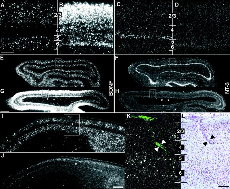
Figure 1.
Alteration of neurotrophin gene expression after kainic acid injections into subplate but not cortical plate. (A–H) Horizontal sections through primary visual cortex at P28 showing in situ hybridization for BDNF and NT-3 mRNA in an unmanipulated case (A and E, C and F) and in a P28 animal that received injections of kainic acid into the subplate at P7 (B and G, D and H). High-magnification photomicrographs in A–D correspond to boxed regions in E–H. BDNF mRNA levels surrounding the injection sites increase throughout the cortical plate after subplate ablation (B); NT-3 mRNA levels in layer 4c decrease below the level of detectability (D). Alterations in mRNA levels for BDNF and NT-3 are present for many millimeters surrounding the two injection sites (asterisks in G and H), whereas the hybridization pattern appears normal for both neurotrophins far posteriorly (right in G and H). (I–L) Neurotrophin mRNA expression does not change after injections of kainic acid directly into layer 4. Sagittal sections through primary visual cortex showing in situ hybridization for BDNF (I and K) or NT-3 (J) mRNA at P28 after an injection of kainic acid into layer 4 of primary visual cortex on P7. The patterns and levels of expression are indistinguishable from unmanipulated or saline-injected animals at the same age. High-magnification photomicrograph in K corresponds to the boxed region in I. Fluorescent beads coinjected with the kainic acid are visible in layer 4 of visual cortex (arrowheads in K), and a scar along the injection track is visible in an adjacent section counterstained with cresyl violet (arrowheads in L). Asterisks (I and J) denote injection sites. Numbers denote cortical layers. Anterior is A–H Left and in I–L Right. Scale bars (A–D, K–L), 200 μm; (E–H, I and L), 2 mm.

